
Mole Removal
With the increased focus on skin care and cancer, many patients show concern about moles. Generally, moles that increase in size, change color, have irregular rather than regular edges, or are associated with bleeding should be examined by an expert for possible mole removal.
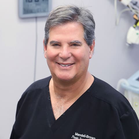
Dr. Mark Mandell-Brown
Dr. Mandell Brown is a nationally recognized, triple board-certified cosmetic surgeon with over 30 years of experience performing facial, breast, and body procedures. Known for his Natural Look™ cosmetic surgery results, Dr. Mandell-Brown has the credentials and aesthetic eye to precisely tailor your procedure and safely achieve the results you desire.

Three mole removal techniques
- Destruction – laser, liquid nitrogen, electrocautery
- Shave Excision – a special razor is used to remove only the mole itself. The skin heals in gradually.
- Full Excision – the mole and adjacent skin are removed and sutures are placed to approximate skin edges.
Not all moles need to be removed. The moles that are suspicious for cancer, irritated by clothing, or are unsightly should be removed. Benefits, risks, and alternatives of each procedure to remove moles can be discussed during your consultation.
Who should treat your facial moles—a family physician, dermatologist, or plastic surgeon? Each physician is influenced by his/her training. The plastic surgeon typically is more concerned with the cosmetic result by virtue of their training. Moles come in many varieties-which should be removed is best determined during a consultation.
Typically, moles that are darkly pigmented, bleed, irritate clothing, are irregular, or increase in size should be removed. Pathologic confirmation with a hospital lab is the only means to determine whether the mole is malignant (cancer) or benign (non-cancer).
Differences in mole removal techniques
Shave Excision — a special surgical blade is used to slice or cut just the mole itself. If the mole is flat or has deeper roots in the skin, removal may leave an indentation. With this technique, the skin heals from the edges. This may take several weeks.
Full Excision — the mole and adjacent skin are removed with a portion of the underlying subcutaneous tissue. The skin edges are then sutured to approximate the skin. This technique leaves a surgical incision line which the surgeon tries to make as small as possible.
Destruction — Electrocautery, laser, and freezing with liquid nitrogen can be utilized to remove the mole. With this technique an indentation at the site is still possible. No specimen is available, however, to send to the lab.
We advise patients who seek improvement at the surgical or biopsy site to consider micropeels, laser resurfacing, or chemical peels after adequate time for healing has occurred.
Areas Served

Medically reviewed by Dr. Mark Mandell-Brown — Updated on Sep 9, 2024